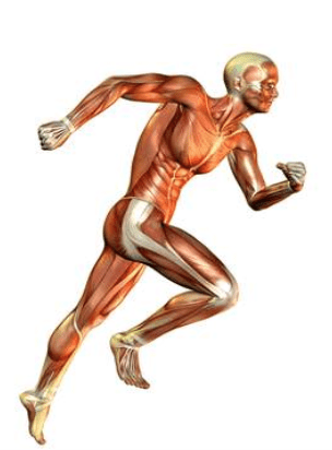
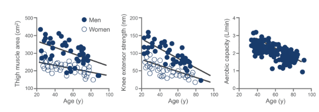
As people age, the following physiological changes are observed in muscle:
1. An increase in fat and connective tissue: Body fat distribution changes with age, with a reduction in subcutaneous fat and an increase in central adiposity. The accumulation of body fat in the abdominal region has been shown to be associated with several adverse health outcomes, such as diabetes, metabolic syndrome, cardiovascular disease (CVD), and all-cause mortality, independently of BMI [1].
2. A decrease in protein synthesis: Ageing is associated with changes in adipose tissue function, which can affect muscle metabolism. Increased levels of TNFα in older individuals can contribute to a pro inflammatory state that affects muscle and fat tissue dynamics [2].
3. A decrease in total muscle cross-sectional area: The amount of tissue inhibitor of metalloproteinases (TIMP) in the dermal layer decreases with ageing. This loss of TIMP may be related to the increase of fat infiltration with ageing, as the balance of matrix metalloproteinases (MMP) and TIMP regulates the boundary between subcutaneous fat and the dermal layer [3].
4. Loss of maximum isometric contraction force: The visceral fat component is more strongly associated with cardiometabolic risk compared to subcutaneous fat, indicating its pathologic role in ageing and muscle function [4].
5. Decreased endurance capacity: Elevated levels of TNFα and other inflammatory markers in older adults can contribute to decreased endurance capacity by promoting inflammation in muscle tissue [5].
In relation to muscle fibres, the faster-contracting type II fibres decrease at a greater rate than type I fibres, leading to a predominance of type I fibres in older individuals. This shift in muscle fibre composition is associated with ageing [6].
It is thought that these age-related changes occur due to:
1. Reduced blood flow to muscles due to decreased capillary density, which limits oxygen availability to exercising muscles. A proinflammatory state in adipose tissue with ageing can contribute to the gradual loss of peripheral fat and affect muscle perfusion [7].
2. A decrease in aerobic enzymes resulting in mitochondrial decay: Ageing is associated with changes in fat metabolism, including increased expression of certain genes in visceral fat that may affect overall energy metabolism [8].
3. Increased mitochondrial DNA deletions and mutations: Research indicates that visceral adipose tissue and liver fat are associated with age-related changes in metabolism, which can impact muscle function [9].
Ageing and Change in Muscle Strength:
It is reported that body muscle mass tends to decrease with ageing [10]. The age- related changes in muscle result in reduced muscle strength. This process begins at age 30, with a subsequent reduction in strength of approximately 8% per decade. The rate of decrease is similar in men and women and affects muscle strength more in the legs than in the arms. By the age of 70, there is a 20–40% decrease in maximal isometric strength, which impacts sustainable walking speed [11].
Sarcopenia:
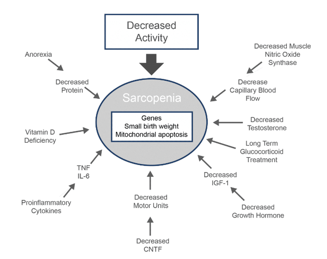
Sarcopenia can be defined as the loss of skeletal muscle mass and function because of ageing [12].
Diagnosis of Sarcopenia:
The European consensus for the diagnosis of sarcopenia requires documentation of criterion 1 and either criterion 2 or 3.
Criterion 1: low muscle mass [13].
Criterion 2: low muscle strength [13].
Criterion 3: low physical performance [13].
References:
1. Mulligan A. , Lentjes M. , Luben R. , Wareham N. , & Khaw K.. Changes in waist circumference and risk of all-cause and cvd mortality: results from the european prospective investigation into cancer in norfolk (epic-norfolk) cohort study. BMC Cardiovascular Disorders 2019;19(1). https://doi.org/10.1186/s12872-019-1223-z
2. Tchkonia T. , Pirtskhalava T. , Thomou T. , Cartwright M. , Wise B. , Καραγιαννίδης Ι. et al.. Increased tnfα and ccaat/enhancer-binding protein homologous protein with aging predispose preadipocytes to resist adipogenesis. American Journal of Physiology-Endocrinology and Metabolism 2007;293(6):E1810-E1819. https://doi.org/10.1152/ajpendo.00295.2007
3. Ezure T. , Amano S. , & Matsuzaki K.. Infiltration of subcutaneous adipose layer into the dermal layer with aging. Skin Research and Technology 2022;28(2):311- 316. https://doi.org/10.1111/srt.13133
4. Kim T. , Lee S. , Yoo J. , Kim D. , Yoo S. , Song H. et al.. The relationship between the regional abdominal adipose tissue distribution and the serum uric acid levels in people with type 2 diabetes mellitus. Diabetology &Amp; Metabolic Syndrome 2012;4(1). https://doi.org/10.1186/1758-5996-4-3
5. Song J. , Farris D. , Ariza P. , Moorjani S. , Varghese M. , Blin M. et al.. Age‐associated adipose tissue inflammation promotes monocyte chemotaxis and enhances atherosclerosis. Aging Cell 2023;22(2). https://doi.org/10.1111/acel.13783
6. Eom J. , Seo J. , y K. , Song S. , Kim G. , & Yang H.. Comparison of chemical composition, quality, and muscle fiber characteristics between cull sows and commercial pigs: the relationship between pork quality based on muscle fiber characteristics. Food Science of Animal Resources 2024;44(1):87- 102. https://doi.org/10.5851/kosfa.2023.e58
7. Caso G. , McNurlan M. , Mileva I. , Zemlyak A. , Mynarcik D. , & Gelato M.. Peripheral fat loss and decline in adipogenesis in older humans. Metabolism 2013;62(3):337-340. https://doi.org/10.1016/j.metabol.2012.08.007
8. Park, S. E., Park, C., Choi, J. M., Chang, E., Rhee, E., Lee, W. Y., … & Soo, B. (2016). Depot-specific changes in fat metabolism with aging in a type 2 diabetic animal model. Plos One, 11(2), e0148141. https://doi.org/10.1371/journal.pone.0148141
9. Losev V. , Lu C. , Senevirathne D. , Inglese P. , Bai W. , King A. et al.. Body fat and human cardiovascular ageing. 2024. https://doi.org/10.1101/2024.06.27.24309526
10. Matsumoto M. , Okada E. , Ichihara D. , Watanabe K. , Chiba K. , Toyama Y. et al.. Changes in the cross-sectional area of deep posterior extensor muscles of the cervical spine after anterior decompression and fusion: 10-year follow-up study using mri. European Spine Journal 2011;21(2):304-308. https://doi.org/10.1007/s00586-011-1978-0
11. Ricciardi C. , Ponsiglione A. , Recenti M. , Amato F. , Gislason M. , Chang M. et al.. Development of soft tissue asymmetry indicators to characterize aging and functional mobility. Frontiers in Bioengineering and Biotechnology 2023;11. https://doi.org/10.3389/fbioe.2023.1282024
12. Sepe A. , Tchkonia T. , Thomou T. , Zamboni M. , & Kirkland J.. Aging and regional differences in fat cell progenitors – a mini-review. Gerontology 2010;57(1):66-75. https://doi.org/10.1159/000279755
13. Gumucio J. and Mendias C.. Atrogin-1, murf-1, and sarcopenia. Endocrine 2012;43(1):12-21. https://doi.org/10.1007/s12020-012-9751-7


